With cutting edge technology, Oregon Heart Center specialists are able to accurately and precisely evaluate your cardiac medical condition.
Below is information provided by the American Heart Association (AHA).
Ambulatory Heart Monitoring
Devices (Pacemaker, ICD, Loop)
Echocardiogram
EKG
Stress Testing
Medication Management
Vascular Ultrasound
EP Study
PET Stress Testing
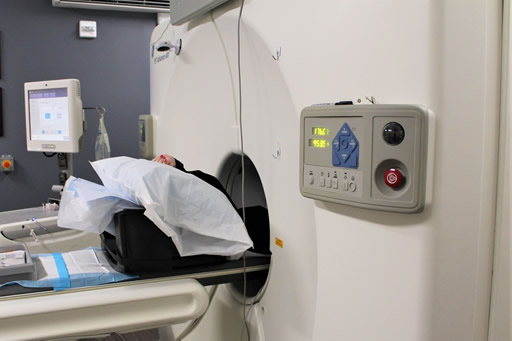
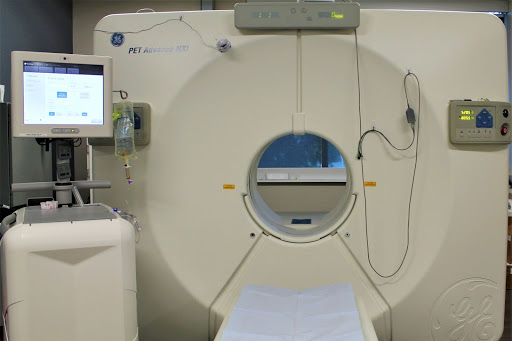
A PET scan of the heart is a noninvasive nuclear imaging test. It uses radioactive tracers (called radionuclides) to produce pictures of your heart. Doctors use cardiac PET scans to diagnose coronary artery disease (CAD) and damage due to a heart attack. PET scans can show healthy and damaged heart muscle. Doctors also use PET scans to help find out if you will benefit from a percutaneous coronary intervention (PCI) such as angioplasty and stenting, coronary artery bypass surgery (CABG) or another procedure.
Quick facts
- PET scans use radioactive material called tracers. Tracers mix with your blood and are taken up by your heart muscle.
- Special positron detectors circle the chest to pick up signals from the tracer. A computer converts the signals into pictures of your heart at work.
- A PET scan shows if your heart is getting enough blood or if blood flow is reduced because of narrowed arteries. It also shows dead cells (scars) from a prior heart attack.
- A PET scan can help in determining if you’ll benefit from a cardiac procedure (PCI) or surgery to restore blood flow. The tracers used for PET scans can help identify injured but still living (viable) heart muscle that might be saved if blood flow is restored.
Why do people have cardiac PET scans?
A PET scan is a very accurate way to diagnose coronary artery disease and detect areas of low blood flow in the heart. PET can also identify dead tissue and injured tissue that’s still living and functioning. If the tissue is viable, you may benefit from a PCI or coronary artery bypass surgery.
What are the risks of cardiac PET?
Cardiac PET is safe for most people. The amount of radiation is small, and your body will get rid of it through your kidneys within about 24 hours. If you’re pregnant or think you might be pregnant, or if you’re a nursing mother, tell your doctor before you have this test. It could harm your baby.
Single Photon Emission Computed Tomography (SPECT)
What is a cardiac SPECT scan?
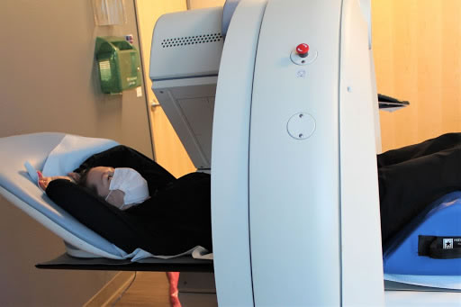
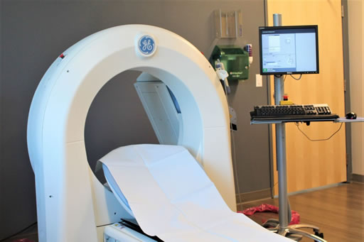
A SPECT scan of the heart is a noninvasive nuclear imaging test. It uses radioactive tracers that are injected into the blood to produce pictures of your heart. Doctors use SPECT to diagnose coronary artery disease and find out if a heart attack has occurred. SPECT can show how well blood is flowing to the heart and how well the heart is working.
Quick facts
- SPECT scans use radioactive material called tracers. The tracers mix with your blood and are taken up by living heart muscle.
- A special “gamma” camera picks up signals from the tracer as it moves around your chest. The tracer’s signals are converted into images by a computer.
- The pictures will help your doctor see if your heart is getting enough blood or if blood flow is reduced because of narrowed arteries.
- A SPECT scan can be used to examine blood flow in your heart at rest and during exercise (called a nuclear stress test). If you can’t exercise, you’ll get a medicine to increase the blood flow in your heart as if you were exercising (called a chemical or pharmacologic stress).
- SPECT scans can also give information about how well your heart is pumping.
Exercise Stress Test
A stress test, sometimes called a treadmill test or exercise test, helps a doctor find out how well your heart handles work. As your body works harder during the test, it requires more oxygen, so the heart must pump more blood. The test can show if the blood supply is reduced in the arteries that supply the heart. It also helps doctors know the kind and level of exercise appropriate for a patient.
A person taking the test:
- is hooked up to equipment to monitor the heart.
- walks slowly in place on a treadmill. Then the speed is increased for a faster pace and the treadmill is tilted to produce the effect of going up a small hill.
- may be asked to breathe into a tube for a couple of minutes.
- can stop the test at any time if needed.
- afterwards will sit or lie down to have their heart and blood pressure checked.
- Heart rate, breathing, blood pressure, electrocardiogram (ECG or EKG), and how tired you feel are monitored during the test.
- Healthy people who take the test are at very little risk. It’s about the same as if they walk fast or jog up a big hill. Medical professionals should be present in case something unusual happens during the test.
Heart rate, breathing, blood pressure, electrocardiogram (ECG or EKG), and how tired you feel are monitored during the test.
Healthy people who take the test are at very little risk. It’s about the same as if they walk fast or jog up a big hill. Medical professionals should be present in case something unusual happens during the test.
Echocardiogram (Echo)
What is an echocardiogram?
An echocardiogram (echo) is a test that uses high frequency sound waves (ultrasound) to make pictures of your heart. The test is also called echocardiography or diagnostic cardiac ultrasound.
Quick facts
- An echo uses sound waves to create pictures of your heart’s chambers, valves, walls and the blood vessels (aorta, arteries, veins) attached to your heart.
- A probe called a transducer is passed over your chest. The probe produces sound waves that bounce off your heart and “echo” back to the probe. These waves are changed into pictures viewed on a video monitor.
- An echo can’t harm you.
Why do people need an echo test?
Your doctor may use an echo test to look at your heart’s structure and check how well your heart functions. The test helps your doctor find out:
- The size and shape of your heart, and the size, thickness and movement of your heart’s walls.
- How your heart moves.
- The heart’s pumping strength.
- If the heart valves are working correctly.
- If blood is leaking backwards through your heart valves (regurgitation).
- If the heart valves are too narrow (stenosis).
- If there is a tumor or infectious growth around your heart valves.
- Blood clots in the chambers of your heart.
- Abnormal holes between the chambers of the heart.
What are the risks?
- An echo can’t harm you.
- An echo doesn’t hurt and has no side effects.
Cardiac Catheterization
What is cardiac catheterization?
Cardiac catheterization (cardiac cath or heart cath) is a procedure to examine how well your heart is working. A thin, hollow tube called a catheter is inserted into a large blood vessel that leads to your heart. View an illustration of cardiac catheterization (link opens in new window).
Quick facts
- Cardiac cath is performed to find out if you have disease of the heart muscle, valves or coronary (heart) arteries.
- During the procedure, the pressure and blood flow in your heart can be measured.
- Coronary angiography (PDF) is done during cardiac catheterization. A contrast dye visible in X-rays is injected through the catheter. X-ray images show the dye as it flows through the heart arteries. This shows where arteries are blocked.
- The chances that problems will develop during cardiac cath are low.
Why do people have cardiac catheterization?
A cardiac cath provides information on how well your heart works, identifies problems and allows for procedures to open blocked arteries. For example, during cardiac cath your doctor may:
- Take X-rays using contrast dye injected through the catheter to look for narrowed or blocked coronary arteries. This is called coronary angiography or coronary arteriography.
- Perform a percutaneous coronary intervention (PCI) such as coronary angioplasty with stenting to open up narrowed or blocked segments of a coronary artery.
- Check the pressure in the four chambers of your heart.
- Take samples of blood to measure the oxygen content in the four chambers of your heart.
- Evaluate the ability of the pumping chambers to contract.
- Look for defects in the valves or chambers of your heart.
- Remove a small piece of heart tissue to examine under a microscope (biopsy).
What are the risks of cardiac catheterization?
Cardiac cath is usually very safe. A small number of people have minor problems. Some develop bruises where the catheter had been inserted (puncture site). The contrast dye that makes the arteries show up on X-rays causes some people to feel sick to their stomachs, get itchy or develop hives.
